Sperry, R. W. (1968). Hemisphere deconnection and unity in conscious awareness. American Psychologist, 23(10), 723.
This is the classic biological psychology study which you will look at for your H167 AS OCR Psychology exam. You will also need this study for your OCR H567 A Level Psychology core studies exam.
Background
The theme of the biological psychology studies in the H167 exam is regions of the brain. This study by Sperry (1968) has the tagline by OCR: Split-brain Study.
What does it mean to have a ‘split-brain’?
The brain is composed of two cerebral hemispheres: the left hemisphere and the right hemisphere. These hemispheres are connected in the brain by the corpus callosum, and other smaller connections, but we need not worry about them here. Having a ‘split-brain’ simply means that the corpus callosum has been severed.
What is the corpus callosum?
The corpus callosum is a large band of white matter (white matter is efficient at sending information), which connects the two hemispheres of the brain.
Why would the corpus callosum be severed?
The corpus callosum may be severed by surgeons in order to reduce the symptoms of epilepsy. In epilepsy, on hemisphere of the brain is usually responsible. Very simply put, when an epileptic episode occurs, there is an electrical storm in one hemisphere of the brain, which then travels across the corpus callosum, causing the entire brain to be affected and then a blackout occurs. By severing the corpus callosum this travelling of the electrical storm cannot occur and thus blackout and epileptic fits cease.
The severing of the corpus callosum is called a commissurotomy.
Difficulties learning Sperry’s Study
One of the most confusing and challenging parts for most students when learning Sperry’s study is the idea of contralateralisation.
The brain is composed of two hemispheres, as mentioned earlier. Most stimuli (sound is more complex) is processed contralaterally. This means that if stimuli enters on the left, for example, the left hand, it is processed in the right hemisphere and visa versa.
For the most part, language is processed in the left hemisphere. (For the most part because this gets complicated, especially when participants’ have different dominant hands, but for the purposes of this study, remember that language, especially spoken language is processed in the left side of the brain).
These two ideas are quite difficult to remember, as it quickly becomes confusing. I suggest testing each other in small groups in order to get to grips with these ideas.
If you want to read more on the work of Roger Sperry, then read: Beyond a World Divided: Human Values in the Brain-mind Science of Roger Sperry
Aim of the Experiment
The aim of Sperry (1968) was to show the independent streams of conscious awareness possessed by each hemisphere and to show how each hemisphere has its own memories.
Method and Design
Sperry (1968) used a quasi experiment in a laboratory with an independent measures design.
The independent variable was whether the individual had a split-brain or not.
There were dependent variable was that individual’s performance on visual and tactile tasks.
Please note, Sperry’s (1968) study did not have a control group. This was because the effects of not having a split-brain were already known. Therefore, there was no need to have a control group.
Further, some have argued that this study was actually a small collection of case studies.
Sample and Sampling Method
11 Participants.
All the participants were epileptics had previously undergone commissurotomies to deal with their severe epileptic convulsions.
The first patient (a man) had his surgery over 5½ years before the study was conducted.
The second patient, a housewife and mother in her 30s had her surgery more than 4 years before the study was conducted.
The other 9 patients had their surgery at varying times but not long before the study was conducted.
Procedure
Visual fields.

We have two visual fields. These visual fields should not be confused with our two eyes. We don’t need to go into the biological factors underlying the visual fields here. The left visual field, as you might guess, is what we can see on our left and the right visual field is what we can see on our right.
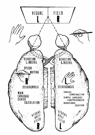
As you can see in the diagram, the information from the right visual field goes to the left hemisphere (see the hand with the pencil).
Visual Tasks
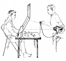
Sperry used a tachistoscope to present visual stimuli to the participants.

The tachistoscope has a focal point in the middle and two areas where stimuli was presented. The participants using the tachistoscope would have one eye covered and were instructed to stare at the focal point.
Information presented to the left of the focal point would be seen in the left visual field which would then travel to the right hemisphere.
Wearing an eye patch and staring at the focal point were controls. These controls ensure that stimuli was presented only to the desired visual field.
All visual stimuli was presented for only 0.1 seconds. This was another control as it is too quick for eye movements to cause visual information to enter both visual fields.
$/? Task. The $/? task had both a ‘$’ and a ‘?’ presented simultaneously (one to the right visual field and the other to the left visual field). Participants were then tested on the stimuli.
Tactile Tasks
Notice in the above image of the tachistoscope there are some objects behind the screen. In the tactile tests the participants would put their hands under the tachistoscope such that they could reach the objects. The participants’ hands were then covered. This was a control to ensure there was no visual stimuli going to either hemisphere and thus could not confound upon the results. Given that the tachistoscope was in between the participants and the objects, the participants could not see the objects. Again this was a control measure to ensure only tactile stimuli was introduced to the participants.
Participants were introduced to objects by an experimenter, who placed them in the participants’ hands.
Objects placed in the right hand of the participant are processed in the left hemisphere.
Objects placed in the left hand of the participant are processed in the right hemisphere.
Results
Visual Tasks
Participants would only recognise stimuli if the stimuli was presented again to the same visual field. If participants were shown stimuli in the right visual field, but then shown the same stimuli to the left visual field, they would claim to have not seen it before.
Information presented to the right visual field (left hemisphere) could be described in speech and writing (with the right hand). If the same information is presented to the left visual field (right hemisphere), the participant insisted he either did not see anything or that there was only a flash of light on the left side, that is, the information could not be described in speech or writing. However the participant could point with his left hand (controlled by the right hemisphere) to a matching picture / object presented among a collection of pictures / objects.
$/? task findings. If different figures were presented simultaneously to different visual field, for example ‘$’ sign to the left visual field and ‘?’ to the right visual field, the participant could draw the ‘$’ sign with his left hand but reported that he had seen a ‘?’.
Tactile Tasks
Objects placed in the right hand (left hemisphere) could be described in speech or writing (with the right hand). If the same objects were placed in the left hand (right hemisphere) participants could only make wild guesses and often seemed unaware they were holding anything.
Objects felt by one hand were only recognised again by the same hand for example objects first sensed by the right hand could not be retrieved by the left.
When two objects were placed simultaneously in each hand and then hidden in a pile of objects, both hands selected their own object and ignored the other hand’s object.
Conclusions
People with split brains have two separate visual inner worlds, each with its own train of visual images.
Split-brain patients have a lack of cross-integration where the second hemisphere does not know what the first hemisphere has been doing.
Split-brain patients seem to have two independent streams of consciousness, each with its own memories, perceptions and impulses ie two minds in one body
Sperry (1968) Evaluation
– Quasi study – as the there was no manipulation by the experimenter, establishing cause and effect becomes more difficult than a traditional experiment.
+ Ethics – as the study used a quasi methodology, Sperry did not need to manipulate anything, which is more ethical than studies which manipulate variables.
– No control group, Sperry did not use a control group, which makes it difficult to truly establish cause and effect. However, it is important to note that in this study a control group was not needed as the results of the tasks for people without split corpus callosums were already known.
– External validity – the external validity in this study may be considered low because the stimuli was selectively delivered to one hemisphere. This does not happen in real life and thus is not representative of the everyday experience of ‘split-brain’ patients. However this selective presentation of stimuli increased the internal validity because there is an increased likelihood that Sperry measured the effects of hemisphere deconnection.
Sperry, R. W. (1968). Hemisphere deconnection and unity in conscious awareness. American Psychologist, 23(10), 723.
Further Reading
Beyond a World Divided: Human Values in the Brain-mind Science of Roger Sperry
Psych Yogi’s Top Ten Psychology Revision Tips for the A* Student

Helped alot for my mock exams tomoz, thanks.
Helpful to prepare the presentation of split-brain study!
Thanks a lot.!
No worries – glad you found it helpful, Daisy